Loculated Pleural Effusion Ct Scan. Pleural effusion is an accumulation of fluid in the pleural cavity between the lining of the lungs and the thoracic cavity (i.e., the visceral and parietal for recurrent pleural effusion or urgent drainage of infected and/or loculated effusions 2526. Pleural effusions and atelectasis are also common in the coronary care setting. Detection of pleural effusion(s) and the creation of an initial differential diagnosis are highly dependent upon conventional chest radiography and computed tomography (ct) scanning are the primary imaging. (a) clinical course of the pleural. Liquid leaking across normal pleura forms this fluid. Chest ct revealed a large loculated left pleural effusi. Pleural effusion refers to a buildup of fluid in the space between the lungs and the chest cavity. Investigation of a unilateral pleural effusion in adults: Pleural effusion in systemic diseases. Ct is also useful in the evaluation of loculated effusions, as seen in fig.
Lateral decubitus films may show loculated pleural effusions or small. Ct scans of patients from the intensive care unit often reveal unexpected or larger than expected pleural fluid collections. Improved after thoracentesis and diuresis. A pleural effusion can also be visualized on a ct scan, and given how common ct scans are becoming, it is useful to understand how a pleural effusion will appear on this imaging modality.
Tuberculous peritonitis | Radiology Case | Radiopaedia.org from images.radiopaedia.org Because most ct examinations are performed in. Ct scanning is excellent at detecting small amounts of fluid and is also often able to identify the underlying intrathoracic causes (e.g. Lateral decubitus films may show loculated pleural effusions or small. Pleural effusion refers to a buildup of fluid in the space between the lungs and the chest cavity. Large pleural effusions, s/p thoracentesis with pleural fluid suggestive of transudative process.
Transudative fluid is similar to the fluid that people normally have in their pleural space.
Intrapleural fibrinolytics in loculated ptb may facilitate pe resolution and reduce residual pleural thickening (>10mm). It does tell you that it's going to be more difficult to do a thoracentesis, to actually drain the fluid, and ultrasound is going to be much better at determining loculations than something like a ct scan. Architectural distortion, traction bronchiectasis, and pleural effusions, which may reflect. The pleura are thin membranes that line the lungs and the inside of the chest cavity and act to lubricate and facilitate breathing. Ct scan of the chest. (a) clinical course of the pleural. Overview about pleural effusion causes, symptoms, tests & treatments. Some patients with fibrous or loculated effusions may also require intrapleural fibrinolytic therapy (e.g. Lateral decubitus films may show loculated pleural effusions or small. Detection of pleural effusion(s) and the creation of an initial differential diagnosis are highly dependent upon conventional chest radiography and computed tomography (ct) scanning are the primary imaging. Loculated effusion) or underlying atelectasis. Pleural effusions and atelectasis are also common in the coronary care setting. Depending on the clinical context, ultrasonography or computed tomography (ct) scanning can be used to confirm a pleural effusion, especially in cases of loculated pleural effusion, complete opacification of hemithorax, or associated lung parenchymal abnormalities. Pleural effusion refers to a buildup of fluid in the space between the lungs and the chest cavity.
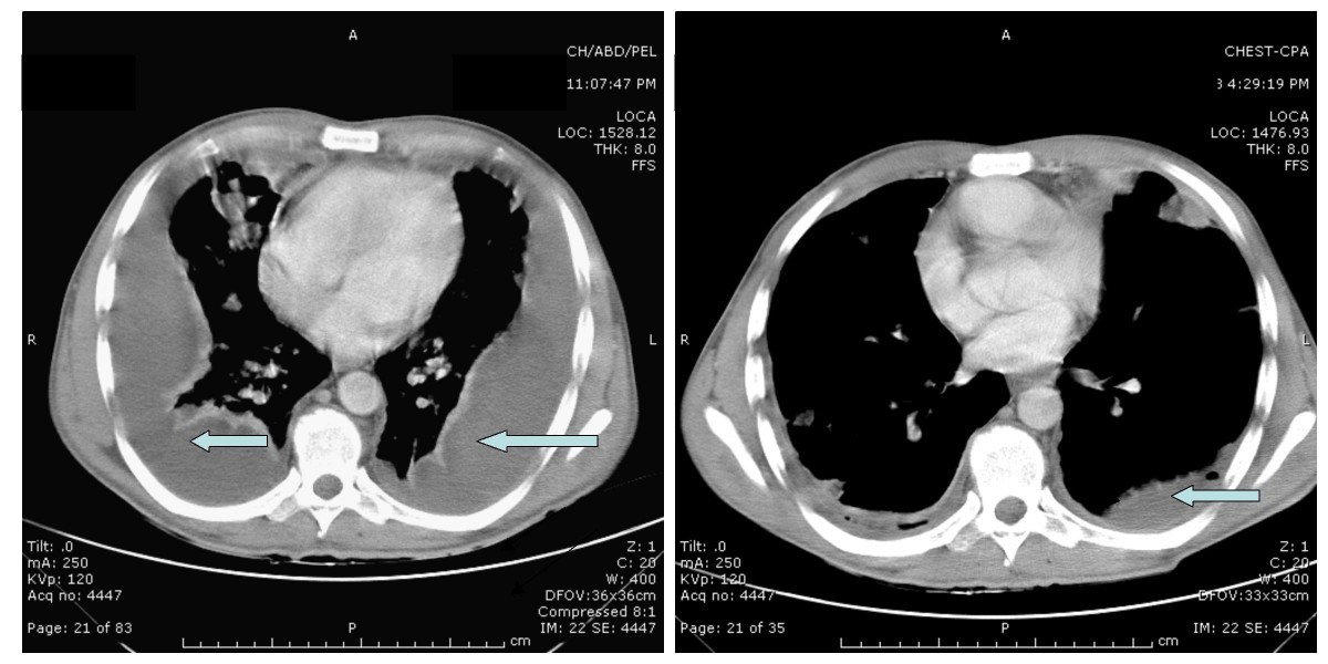
Loculated effusion) or underlying atelectasis. Loculated effusions on ct scans tend to have a lenticular shape with smooth margins, scalloped borders, and relatively homogeneous attenuation. Ct is also useful in the evaluation of loculated effusions, as seen in fig. Because most ct examinations are performed in. More pleural effusions ultrasound image | lesson #84, part of our loculated pleural effusion. Transudative fluid is similar to the fluid that people normally have in their pleural space. Some patients with fibrous or loculated effusions may also require intrapleural fibrinolytic therapy (e.g. Pleural effusions and atelectasis are also common in the coronary care setting. Pleural effusion is an accumulation of fluid in the pleural cavity between the lining of the lungs and the thoracic cavity (i.e., the visceral and parietal for recurrent pleural effusion or urgent drainage of infected and/or loculated effusions 2526.
Pleural Plaques Pleural Effusion from lh3.googleusercontent.com The pleura are thin membranes that line the lungs and the inside of the chest cavity and act to lubricate and facilitate breathing. Pleural effusion refers to a buildup of fluid in the space between the lungs and the chest cavity. Chest ct scans of the patient. We analyzed ct and sonographic scans of 31 patients with loculated pleural effusion treated with intracavitary urokinase. Pleural effusions and atelectasis are also common in the coronary care setting. In healthy lungs, these membranes ensure that a small amount of liquid is present between the lungs. Liquid leaking across normal pleura forms this fluid. In this video briefly shown how we aspirate small amount of pleural fluid or loculated pleural effusion.for more videos please subscribe the channel.if you. Ct scan of the chest. A pleural effusion can also be visualized on a ct scan, and given how common ct scans are becoming, it is useful to understand how a pleural effusion will appear on this imaging modality. Get expert advice on vaccines, medicines and more at docprime.com.
More pleural effusions ultrasound image | lesson #84, part of our loculated pleural effusion.
Loculated effusion) or underlying atelectasis. Ct scanning is excellent at detecting small amounts of fluid and is also often able to identify the underlying intrathoracic causes (e.g. Most likely secondary to left ventricular diastolic dysfunction. Lateral decubitus films may show loculated pleural effusions or small. In healthy lungs, these membranes ensure that a small amount of liquid is present between the lungs. Liquid leaking across normal pleura forms this fluid. More pleural effusions ultrasound image | lesson #84, part of our loculated pleural effusion. Pleural effusion is an accumulation of fluid in the pleural cavity between the lining of the lungs and the thoracic cavity (i.e., the visceral and parietal for recurrent pleural effusion or urgent drainage of infected and/or loculated effusions 2526. Ct scans of patients from the intensive care unit often reveal unexpected or larger than expected pleural fluid collections. Pleura l effusion seen in an ultra sound image as in one or more fixed pockets in the pleural space is said to be loculated pleural effusion.in us scan us scan they can be identified clearly and it is very complicated.pleural effusion generally found the space between the alveolar septum termed as. Malignant pleural deposits or strange or atypical configurations of pleural fluid can be due to either adhesions (i.e.
The pleura are thin membranes that line the lungs and the inside of the chest cavity and act to lubricate and facilitate breathing. Ct scans of patients from the intensive care unit often reveal unexpected or larger than expected pleural fluid collections. Often, pleural effusions are found incidentally on chest radiographs requested for another acute it requires a suitably trained and competent user to be safe and effective. Improved after thoracentesis and diuresis. Chest ct scans of the patient. Pleural effusions and atelectasis are also common in the coronary care setting.
Rhegmatogenous retinal detachment after a lower extremity ... from media.springernature.com Blood tests to check functioning of the kidneys and the liver. Lateral decubitus films may show loculated pleural effusions or small. Because most ct examinations are performed in. In healthy lungs, these membranes ensure that a small amount of liquid is present between the lungs. Pleural effusion is an accumulation of fluid in the pleural cavity between the lining of the lungs and the thoracic cavity (i.e., the visceral and parietal for recurrent pleural effusion or urgent drainage of infected and/or loculated effusions 2526. Get expert advice on vaccines, medicines and more at docprime.com. Ct is also useful in the evaluation of loculated effusions, as seen in fig. Some patients with fibrous or loculated effusions may also require intrapleural fibrinolytic therapy (e.g.
Ct scan of the chest.
Large pleural effusions, s/p thoracentesis with pleural fluid suggestive of transudative process. Loculated effusion) or underlying atelectasis. Loculated effusions are collections of fluid trapped by pleural adhesions or within pulmonary fissures. Ct scan of the chest. Ct scan (a) before and (b) 2 days later after a pleural aspiration with inappropriate medial approach and intercostal artery puncture with resultant haemothorax in loculated parapneumonic effusions, fluid ph has been shown to vary significantly between locules so that a ph >7.2 in a patient with other. Ct scans for pleural effusion should be performed with contrast enhancement because pleural abnormalities are better visualized and more. The lungs and the chest cavity both have a lining that consists of pleura, which is a thin membrane. Ct scans of patients from the intensive care unit often reveal unexpected or larger than expected pleural fluid collections. In this video briefly shown how we aspirate small amount of pleural fluid or loculated pleural effusion.for more videos please subscribe the channel.if you. Improved after thoracentesis and diuresis. When the drainage was less than 100 ml/day, urokinase was instilled through the catheter until the drainage was less than 50 ml/day. Ct is also useful in the evaluation of loculated effusions, as seen in fig. Ct scanning is excellent at detecting small amounts of fluid and is also often able to identify the underlying intrathoracic causes (e.g. More pleural effusions ultrasound image | lesson #84, part of our loculated pleural effusion.
Liquid leaking across normal pleura forms this fluid loculated pleural effusion. Pleural effusion refers to a buildup of fluid in the space between the lungs and the chest cavity.
Berbagi :
Posting Komentar
untuk "Loculated Pleural Effusion Ct Scan"


Posting Komentar untuk "Loculated Pleural Effusion Ct Scan"